|
|
|
| A Comparative Evaluation Of The Fracture Resistance Of Teeth Restored With Glass Fiber Post And Biological Dentin Post Cemented With Adhesive Resin: An In - Vitro Study |
Aparna Palekar 1 , Vijay Mantri 2 , Shilpa N. Kamble 3 , Syed Gufran Ali 4 , Shaliputra P.Magar 5
1 Professor & Head, Dept. Conservative Dentistry And Endodontics - Modern Dental College And Research Centre, Gandhi Nagar, Indore (M.P.)
2 Professor, Dept. Of Conservative Dentistry And Endodontics - Modern Dental College And Research Centre, Gandhi Nagar, Indore (M.P.)
3 PG Student, Dept. Conservative Dentistry And Endodontics - Modern Dental College And Research Centre, Gandhi Nagar, Indore (M.P.)
4 Reader , Dept. Of Conservative Dentistry And Endodontics - Modern Dental College And Research Centre, Gandhi Nagar, Indore (M.P.)
5 Senior Lecturer , Dept. Oral Medicine And Radiology - Shro Aurbindo College Of Dentistry Indore
|
| Address For Correspondence |
Dr. Aparna Palekar, Professor & Head,
Dept. of Conservative Dentistry and Endodontics
Modern Dental College and Research Centre,
Gandhi Nagar, Indore (M.P.)
Phone no. : 07354678260
Email : aparnapalekar@hotmail.com |
| Abstract |
| Aim: The aim of this study was to evaluate and compare the fracture resistance of endodontically treated permanent maxillary central incisors restored with glass fiber post biological dentin post cemented with adhesive resin.
Materials and Methods: Root canal treatment was performed on all 80 maxillary central incisors and samples were divided into four groups of 20 each. Group 1: Restored as a positive control group without post space preparation and post cementation. Group 2: Restored with fiber post cemented with adhesive resin Group 3: Restored with biological dentin posts cemented with adhesive resin and Group 4: Restored as negative control group with post space preparation but no post placement. The teeth were loaded at 135° angle to their long axis after core build-up and the failure loads were recorded.
Results: One-way Analysis of Variance (ANOVA) and Bonferroni multiple comparisons revealed a significant difference among test groups with the positive control group showing the highest fracture resistance, followed by the dentin post group and lastly the FRC post group. The negative control group showed the least fracture resistance among the groups.
Conclusion: Teeth restored with dentin posts exhibited better fracture resistance than those restored with FRC posts. |
|
| Keywords |
| Dentin Post, Biological post, Flexural strength |
|
| Full Text |
Introduction
Fracture of root filled teeth can be prevented by their proper restoration and reinforcement. The amount of remaining tooth structure is an important consideration for treatment planning. In case of badly broken down teeth where little tooth structure remains the root canal space is utilized for support of the crown restoration[1]. The resultant post and core provides the required retention and resistance form for the final restoration. The post and core equally distributes the torquing forces within the radicular dentin to the supporting tissues. It disperses the forces along the root and provides retention for the core that replaces the lost coronal tooth structure. [2],[3]
The fracture resistance of an endodontically treated tooth can be determined by the amount of remaining tooth structure. The post material and design plays a significant role in determining strength. Different posts have different physical properties. The post material should ideally exhibit physical properties like modulus of elasticity, compressive strength and thermal expansion as well as aesthetics similar to that of dentin. It should also bond predictably to root dentin.[4] The only material that fulfills all these requirements is none other than dentin itself. Biological posts made of root dentin exhibit properties similar to the tooth.[5] Hence its use as a post should be investigated.
The aim of this ex vivo study was to evaluate and compare the fracture resistance of endodontically treated maxillary central incisors restored with prefabricated fiber reinforced composite (FRC) posts and experimental dentin posts.
Materials & Method:
Eighty freshly extracted maxillary central incisors with inclusion criteria of completely formed roots of similar sizes and exclusion criteria of absence of caries, visible fracture lines or cracks were selected for the study. Root canal treatment was performed on all the 80 specimens. Obturation was carried out by the cold lateral condensation method using a 40-size gutta percha (Dentsply) as master cone and AH-plus (Dentsply-Kronstaz, Germany), a non-eugenol endodontic sealer. The crown of each tooth was reduced to a height of 2 mm above the cemento-enamel junction in order to simulate the clinical situation of a reduced tooth structure so that the resistance to fracture of the post system used would be more relevant clinically. Post space was prepared for all the 60 specimens (20 specimens kept as a control group without post and post space preparation). A post space of depth 10 mm was standardized from the cut tooth surface that was taken as the reference point. A thin coat of polyvinylsiloxane (Aquasil ultra LV, Dentsply, Germany) was painted on the root surfaces of all teeth to within 1 mm of the CEJ, to simulate the effect of periodontal ligament. Samples were embedded obliquely in triangular shaped acrylic blocks of 3×3×4 cm. During the procedure, samples were kept in distilled water to provide moist environment to avoid dehydration of the dental tissues. The prepared teeth were divided into four groups of twenty specimens each.
Grouping of teeth and post insertion:
Group 1: Twenty teeth which were obturated but with no post space preparation. The restoration of access openings done was with composite restoration (3M ESPE). (Positive Control group)
Group 2: Twenty teeth restored with prefabricated glass fiber-reinforced composite tapered posts (REFORPOST, Angelus)
Group 3: Twenty teeth restored with biological dentin posts.
Group 4: Twenty teeth in which post space preparation done without any post cementation and without restoration. (Negative Control group)
The posts were cemented using adhesive dual cure resins (Panavia F, Kuraray Co. Ltd., Japan). The core was fabricated with composite resin (3M ESPE FiltekTM Z 350 XT) for all groups.
Preparation of experimental dentin posts:
Ten healthy maxillary canines freshly extracted for periodontal reasons were selected. Each tooth was sectioned longitudinally (mesiodistally) into two halves along the root canal. Template was made of Pre-fabricated glass fiber posts which were selected for the study on putty impression material. Cylindrical dentin blocks were prepared out of each section using diamond drills under intense water cooling and were then subjected to generate twenty dentin posts of standardized shape and dimensions (12 mm length, 1.6 mm diameter) similar to FRC posts. (Fig : 1)
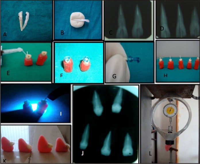 | Figure 1
 |
Investigation of fracture resistance
The samples were subjected to thermocycling (5000 cycles between 500C to 550C with a dwell time of 30 sec at each temperature) and stored in distilled water for 24 hrs at 370C in a humidor (100% relative humidity) to simulate conditions in the oral cavity prior to the fracture test. A compressive load was applied using universal loading machine at CEJ on the palatal aspect, at an angle of 135o to the long axis of the tooth at a crosshead speed of 5 mm/min. The load at fracture was measured and the mean was calculated using statistical analysis of Post Hoc Tests and Bonferroni testand the significance among the four groups was analyzed. The failure threshold was defined as the point at which the loading force reached the maximum value for fracturing the root, post or core. Mean failure load values were calculated for all groups. Data was analyzed using SPSS 14 software.
The descriptive statistics like mean and standard deviation of the fracture of positive control group, resistance of teeth restored with fibre reinforced composite (FRC) posts, experimental dentin posts and negative control group were identified. Prevalence of an outcome variable along with 95% confidence interval for mean was calculated.
Results
Positive Control group (Group 1) showed the maximum mean failure load value at 147.04 Kg, followed by the dentin post group (Group 3) and FRC post group (Group 2) at 116.20 Kg and 97.10 Kg respectively. Negative control group (Group 4) showed the least mean failure load at 68.20 Kg [Table 1]. Oneway ANOVA showed significant difference (P < 0.001) among the test groups [Table 2].
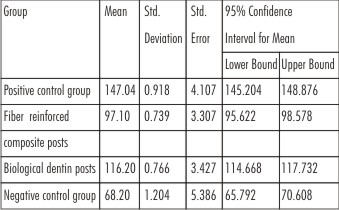 | Table 1: Mean failure load values for fracture resistance of tooth (in Kg)
|
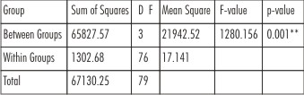 | Table 2: One-way Analysis of Variance (ANOVA)
|
Post Hoc Testsmultiple comparison test [Table 3] revealed a significantly higher fracture resistance for the control group. All the groups differed significantly from each other. Group 2 had a significantly lower fracture resistance than Groups 1 and 3 whereas group 4 had least fracture resistance among groups.
 | Table 3: Multiple Comparisons by Post Hoc Test
|
Discussion
The aim of this study was to evaluate the fracture resistance of teeth restored with glass fiber post and dentin post. The teeth selected for this in-vitro study were maxillary central incisors with the mean size of roots 13.45±0.22 mm in length, 6.35±0.12 mm in mesiodistal, and 6.95 ±0.25 mm in buccopalatal width. Thus standardization of the samples was maintained.
The sectioned roots were not embedded directly into the resin blocks. A thin layer of polyvinyl siloxane covered the roots. As this has modulus of elasticity very similar to natural periodontal ligament it simulated the same. The external reinforcement of embedded roots by the rigid acrylic resin was avoided. [6]
The standardization for post space preparation was achieved by use of calibrated low-speed drill provided by the manufacturer of FRC post to maintain the dimensions of the dentin post and post space same as that of FRC post. To eliminate any procedural technique sensitivity, a custom transparent matrix was used to standardize the dimensions of core build-ups. Dual-cure cement was used for the cementation of the post and for core build-up. This dual-cure resin cement has combined properties of both photo curing (sufficient time and control for proper seating of the post into the canal) and auto curing (polymerization without the influence of post space depth) systems. Moreover, this cement does not require any surface pre-treatment of the substrate such as silanation or etching. [7]
The compressive load was applied at a speed of 5 mm/min at an angle of 135°to the long axis of experimental teeth. This angle reflects the positions, contacts and loading characteristics of upper anterior teeth in Class I occlusion.[8] Guzy and Nicolls reported that for incisors, a loading angle of 130-135º is chosen to simulate a contact angle found in Class I occlusion between maxillary and mandibular anterior teeth[9]
The mean failure load value of the positive control group in our study was more than the experimental dentin post group because of more amount of remaining tooth structure.
Negative control group (tooth with post space prepared but without post cementation) showed the least fracture resistance among groups. This may be because of the hollow space, which did not allow even distribution of the load applied thus leading to fracture of the tooth. It has been suggested that remaining dentin thickness is a critical factor in the resistance of the dentin/root restorative complex during function. [10]
The determination of fracture resistance is of great importance after post cementation as, if the root fractures the tooth is invariably lost. A rigid post has a high modulus of elasticity which causes more stress to root dentin leading to irreparable damage.[10] In an attempt to reduce stresses on the root structure a post having modulus of elasticity similar to the dentin should be used. Carbon fiber, glass fiber posts having modulus of elasticity nearly identical to the dentin have been reported to cause less stress in the tooth and fewer root fractures[11], [12]. This results in a homogeneous unit causing reduction of stresses on the root.[13] A similar homogeneous unit is formed with a dentin post that results in uniform stress distribution. As the property of root dentin and dentin post are similar, both the units flex in a similar manner. The dentin post acts as a shock absorber, transmitting only a fraction of the stresses to the dentinal walls.[14] Gianluca Plotino et al in 2007 reported that flexural strength of FRC post and metal posts was respectively four and seven times higher than root dentin.[15] Metal posts have a high modulus of elasticity (110 GPa), which means that they are stiff and able to withstand forces without distortion. When a force is placed on a tooth containing a stiff post, it is transmitted to the less rigid root dentin, and concentrates at the apex of the post. Stress concentration in the post/root complex increases the chances of fracture. To overcome the concerns about unfavourable stress distribution generated by metal posts, fiber-reinforced composite resin posts were introduced in 1990, with the aim of providing more elastic support to the core.[16] The reduced stress transfer to tooth structure was claimed to reduce the likelihood of root fracture.Posts made of materials with a modulus of elasticity similar to dentin are more resilient, absorb more impact force, and distribute the forces better than stiffer posts.[17]
The failure of the FRC post group in this study may be attributed to the difference in the biomechanical properties between the FRC post and the root dentin. The modulus of elasticity of glass fiber posts is ~ 40 GPa whereas the modulus of elasticity of root dentin is ~ 14.2 GPa and of core material is ~ 13.5 GPa. This difference might create stresses at different interfaces and the possibility of post separation and failure. An added reason for failure at the post cement interface is the presence of interfacial gaps. Moreover, since the resin chemistry of the epoxy resin based posts and methacrylate-based adhesive resin differs completely, the adhesion achieved may not be reliable.[18] In the present study, teeth restored with solid dentin posts exhibited higher fracture resistance than those restored with FRC posts This is in accordance with a study conducted by Craig et al.[19] This can be explained on the basis that the Physiomechanical properties of dentin post are similar to dentin causing uniform stress distribution.[20] The potential advantages of dentin or biological post are: (1) does not promote dentin stress, (2) preserves the internal dentin walls of the root canal, (3) presents total biocompatibility and adapts to conduct configuration, favouring greater tooth strength and greater retention of these posts as compared to premanufactured posts, (4) presents resilience comparable to the original tooth, and (5) offers excellent adhesion to the tooth structure and composite resin and at a low cost. [21]
The limitations to the use of natural biological post made from extracted teeth are (1) Difficulty of finding teeth with a similar color and shape as that of the destroyed tooth, (2) Patient may refuse to accept a tooth fragment obtained from another patient, which prevents the execution of the restoration. [22]
Conclusion
Within the limitations of this study, it can be concluded that:
Teeth restored with dentin posts exhibit better fracture resistance than those restored with FRC posts. This pilot study opens an introductory gate in support of the clinical implications of dentin posts. Dentin post may be a successful alternative to currently available post materials. However, further in vivo trials are required in this direction.
References
1. Fernandes AS, Dessai GS. Factors affecting the fracture resistance of post core reconstructed teeth: A review. Int J Prosthodont 2001; 14:355-63.
2. Schwartz RS, Robbins JW. Post placement and restoration of endodontically treated teeth: A literature review. J Endod 2004; 30:289-301.
3. Prabeesh PadmanabhanA comparative evaluation of the fracture resistance of three Different pre-fabricated posts in endodontically treated Teeth: an in vitro study Journal of Conservative Dentistry | Jul-Sep 2010 | Vol 13 | Issue 3
4. Cheung W. A review of the management of endodontically treated teeth. Post, core and the final restoration. J Am Dent Assoc 2005; 136:611-9.
5. Sirimai S, Riis DN, Morgano SM. An in vitro study of the fracture resistance and the incidence of vertical root fracture of pulpless teeth restored with six post-and-core systems. J Prosthet Dent. 1999;81:262–269
6. Necdet Adanira, Sema Bellib, DDS, PhDEvaluation of Different Post Lengths’ Effect on Fracture Resistance of a Glass Fiber Post System. Eur J Dent 2008; 2:23-28
7. Ceballos L, Garrido MA, Fuentes V, Rodriguez J. Mechanical characterization of resin cements used for luting fiber posts by nanoindentation. Dent Mater 2007; 23:100-5.
8. Martínez-Insua A, da Silva L, Rilo B, Santana U. Comparison of the fracture strength of pulpless teeth restored with a cast post and core or carbon fiber post with a composite core. J Prosthet Dent 1998; 80:527-32
9. Guzy GE, Nicholls JI. In vitro comparison of intact endodontically treated teeth with and without endo-post reinforcement. J Prosthet Dent 1979; 42:39-44.
10. Maccari PC, Conceição EN, Nunes MF. Fracture resistance of endodontically treated teeth restored with three different prefabricated esthetic posts. J Esthet Restor Dent 2003; 15; 25-31.
11. Ho MH, Lee SY, Chen HH, Lee MC. Three dimensional finite element analysis of the effects of posts on stress distribution in dentin. J Prosthet Dent 1994; 72:367-72.
12. Lippo V.J. Lassila, Johanna Tanner Flexural properties of fiber reinforced root canal postsDental Materials (2004) 20, 29–36.
13. Amandeep Kaur, Meena N, Shubhashini N A comparative study of intra canal stress pattern in endodontically treated teeth with average sized canal diameter and reinforced wide canals with three different post systems using finite element analysisJ Conserv Dent | Jan-Mar 2010 | Vol 13 | Issue 1
14. Ambica Kathuria et al Exvivo fracture resistance of endodontically treated maxillary central incisors restored with fiber-reinforced composite posts and experimental dentin posts J. of Cons Dent. Oct-Dec 2011/Vol 14/ Issue 4
15. Gianluca Plotino et al. Flexural properties of endodontic posts and human root dentin Dent. Mat 2007; 23, 1129-1135
16. Marga Ree, DDS, MSca, Richard S. SchwartzThe Endo-Restorative Interface: Current Concepts Dent Clin N Am 54 (2010) 345–374
17. Tay FR, Loushine RJ, LambrechtsP, et al. Geometric factors affecting dentin bonding in root canals: a theoretical modeling approach. J Endod 2005; 31(8):584–9.
18. Lawrence W. Stockton Factors affecting retention of post system: A literature review J. Prostho Dent 1999; 81:380-5
19. Craig RG, Peyton FA. Elastic and mechanical properties of human dentin. J Dent Res 1958; 37:710-8.
20. Newman MP, Yaman P, Dennison J, Rafter M, Billy E. Fracture resistance of endodontically treated teeth restored with composite posts. J Prosthet Dent 2003; 89:360-7.
21. El Mowafy OM, Watts DC. Fracture toughness of human dentin. J Dent Res 1986; 65:677-81
22. Patricia Correa et al “Biological Restoration” Root Canal and Coronal Reconstruction J. Esthet Restor Dent 22:168-178,2010
|
|
|
|
|
|
|